Wisdom Teeth Removal and Nerve Concerns Explained
Wisdom teeth removal is routine oral surgery, but the word "routine" can hide the part patients worry about most: nerves. That concern is reasonable. Lower wisdom teeth sit in one of the most anatomically crowded areas of the mouth, close to structures that control feeling in the lip, chin, gums, and tongue. When a dentist or oral surgeon recommends removing them, many patients hear two thoughts at once. The first is relief that a painful or troublesome tooth can be dealt with. The second is fear about numbness, tingling, or permanent change.
Most of the time, those fears do not become reality. Even when lower wisdom teeth are close to a nerve, careful planning, good imaging, and sound surgical judgment usually lead to a smooth outcome. Still, nerve injury is not a made-up risk, and it deserves a clear explanation. Patients do better when they understand what nerves are involved, how surgeons evaluate risk, what warning signs matter, and how recovery usually unfolds.
Why lower wisdom teeth raise the nerve question
Upper wisdom teeth can create their own problems, especially with the sinuses, but nerve concerns are much more often discussed with lower third molars. The reason is anatomy. The roots of a lower wisdom tooth may develop very close to the inferior alveolar nerve, which runs inside the jawbone. That nerve supplies feeling to the lower lip, chin, and lower teeth on that side. Nearby, another nerve called the lingual nerve runs closer to the inner side of the lower jaw and helps carry feeling, including some taste-related sensation, from part of the tongue.
When people talk about numbness after wisdom teeth removal, they are usually talking about one of these nerves. If the inferior alveolar nerve is irritated, a patient might notice altered feeling in the lower lip or chin. If the lingual nerve is affected, the tongue may feel numb, tingly, or strangely thick on one side. These changes can be temporary and improve as the nerve recovers, but they are unnerving when they happen.
What matters here is proximity, not just the presence of wisdom teeth. Some impacted wisdom teeth are far enough from the nerve canal that the risk is very low. Others lie against it, wrap around it, or sit in a position where the roots and nerve appear superimposed on imaging. That is where an experienced surgeon earns trust, not by promising zero risk, but by explaining the real level of risk and choosing the safest path.
The nerves involved, in plain terms
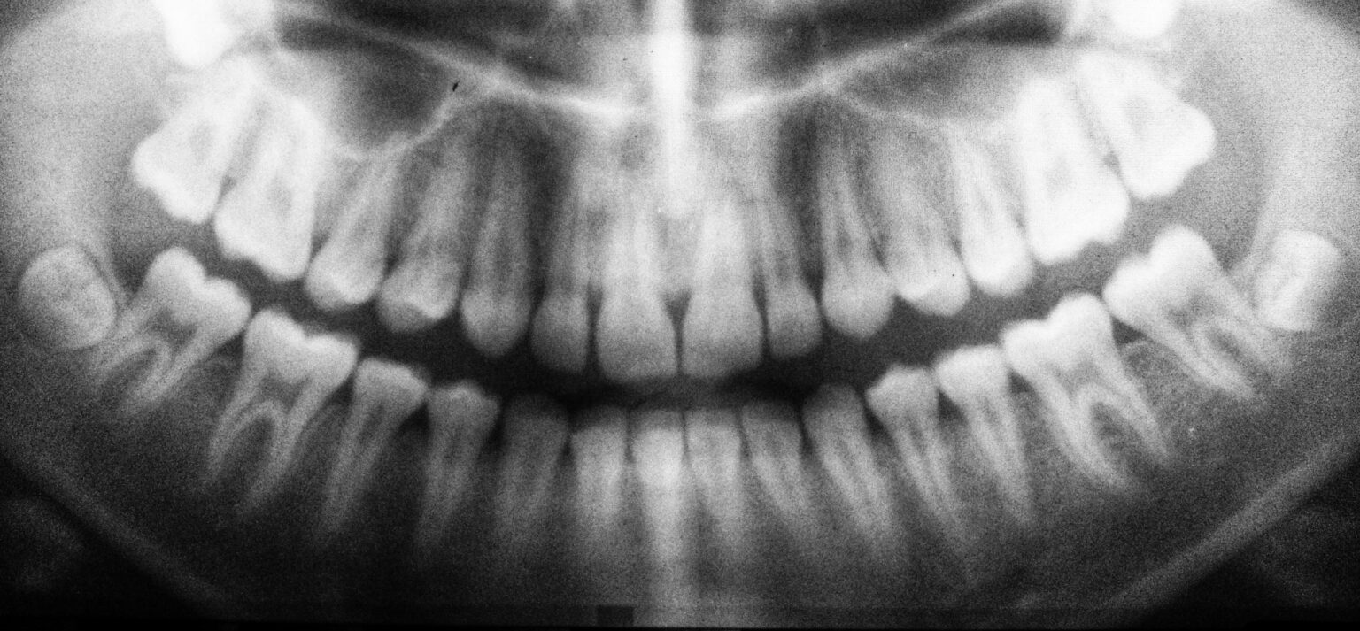
The inferior alveolar nerve sits inside a bony tunnel called the mandibular canal. On an X-ray, that canal often appears as a dark band outlined by thin white borders, running below the roots of lower teeth. If a wisdom tooth root overlaps or distorts that canal, the surgeon pays close attention. Pressure, traction, or surgical manipulation near this area can irritate the nerve.
The lingual nerve is trickier in a different way. It is not encased in bone in the same manner. Instead, it runs through soft tissue along the inside of the lower jaw, near where a surgeon may reflect tissue during extraction. Its exact position can vary slightly from person to person. That variability is one reason surgeons are deliberate with instruments and tissue handling on the tongue side of the lower wisdom tooth.
Patients often ask whether nerve injury means the nerve has been "cut." Usually, no. The more common scenario is temporary nerve bruising or stretching, sometimes called neurapraxia in clinical settings. That can produce numbness, tingling, or altered sensation for days, weeks, or months, then gradually improve. More severe injuries are less common. Permanent sensory change is possible, but it is far less common than temporary disturbance.
How dentists and oral surgeons estimate risk before surgery
A lot of anxiety can be reduced before the day of surgery by doing proper imaging and reading it well. Most patients start with a panoramic X-ray. It gives a broad view of the jaws, tooth position, sinus relationship, and the general course of the lower nerve canal. A skilled clinician can often spot features that suggest the roots are close to the nerve.
Certain radiographic signs tend to raise concern. The canal may appear narrowed, diverted, or darkened where it crosses the roots. The roots themselves may look interrupted or unusually hooked around the canal. None of these signs guarantee a problem, but they can justify closer study.
That is where three-dimensional imaging, usually a cone beam CT scan, becomes valuable. A CBCT does not make surgery risk-free, but it can answer questions a flat X-ray cannot. It shows whether the nerve canal lies to the cheek side, tongue side, between the roots, or just underneath them. That distinction matters. I have seen cases where a panoramic film looked alarming, yet the CBCT showed a thin but safe bony separation. I have also seen the reverse, where a tooth looked manageable on a simple film and the 3D scan revealed a much tighter relationship than expected.
Good planning is not just about deciding whether to remove the tooth. It is also about deciding when, how, and sometimes whether to modify the approach.
When removal is straightforward, and when it is not
Not every impacted wisdom tooth should come out immediately. Some are fully buried, asymptomatic, and stable. Others repeatedly trap food, inflame the gum behind the second molar, damage the neighboring tooth, or develop cystic changes. The decision is rarely one-size-fits-all.
Age matters. Younger patients often have roots that are not fully formed, and the surrounding bone tends to be less dense. That can make extraction easier and may reduce certain risks. In older adults, roots are usually fully developed, bone can be denser, and healing can be slower. That does not mean older patients should avoid surgery, only that the calculus can change.
The position of the tooth matters just as much. A partially erupted lower wisdom tooth that causes repeated infections may be less risky to remove than a completely buried tooth whose roots wrap around the nerve canal. A clinician with judgment weighs symptoms, future risk, anatomy, and the patient's overall health rather than treating every third molar the same way.
Coronectomy, the lesser-known option
One of the most useful developments in high-risk lower wisdom teeth management is the coronectomy. In a coronectomy, the crown of the tooth is removed, but the roots are intentionally left behind when they are judged too close to the inferior alveolar nerve. The goal is to eliminate the part of the tooth causing trouble while lowering the chance of nerve injury.
This option is not appropriate for every case. If the tooth has active root infection, severe mobility, or certain patterns of decay, leaving roots may not be wise. But in properly selected patients, coronectomy can be a very sensible compromise. Patients are sometimes surprised by the idea of intentionally leaving part of a tooth in place, yet it is grounded in a practical principle: do not create more harm than the disease itself.
The roots left after coronectomy often remain quiet. Sometimes they migrate slightly over time, moving away from the nerve and becoming easier to remove later if ever needed. Not every surgeon offers coronectomy, and not every case qualifies, but when the nerve risk is substantial, it deserves discussion.
What nerve injury actually feels like
Patients tend to imagine complete deadness, but nerve symptoms can vary. Some people describe a patch of numbness, as if local anesthetic never fully wore off. Others notice tingling, burning, crawling sensations, or a feeling that the lip or tongue is swollen when it is not. Speech may feel odd for a while, especially with tongue involvement. Eating and drinking can feel clumsy. A few patients accidentally bite the lip or tongue because normal feedback is dulled.
These symptoms can be disturbing even when they are improving. Nerves often recover slowly and imperfectly at first. A patient may move from numbness to pins-and-needles, then to oversensitivity, then to near-normal feeling. That uneven pattern does not necessarily mean something is wrong. It is often how nerve healing behaves.
One practical point is easy to miss: lingering numbness immediately after surgery is not always a red flag. Long-acting local anesthetic can keep tissues numb for many hours. The timing matters. If sensation returns gradually over the same day, that is expected. If there is clear altered sensation beyond the expected anesthetic window, the surgeon should know.
The real numbers, and why they vary
Patients often ask for a percentage risk. That is fair, but the answer depends on the tooth, the imaging, the technique, and the operator's experience. Reported rates of temporary sensory disturbance after lower wisdom tooth removal vary across studies, often sitting in the low single digits, though higher-risk anatomy can push the number up. Permanent nerve injury is much less common than temporary nerve disturbance.
Those ranges vary because studies define injury differently and follow patients for different lengths of time. A brief tingling that resolves in two weeks and a six-month numb patch do not carry the same weight, yet some broad discussions lump them together. That is why a personalized risk estimate from the treating surgeon means more than a generic internet number.
A responsible clinician does not quote statistics as if they erase anatomy. If your roots appear clearly separated from the canal, the conversation will sound very different from one involving intimate contact with the nerve. Good consent is specific.
Surgical technique matters more than many patients realize
Two surgeons can face the same tooth and not perform the same operation in the same way. Technique affects force, visibility, bone removal, and tissue handling. Careful flap design, controlled bone removal, sectioning the tooth into pieces instead of levering aggressively, and avoiding unnecessary traction all help reduce risk.
The best oral surgeons are rarely the most dramatic. They are methodical. They stop when the tissue is not behaving as expected. Wisdom Teeth Removal They change the plan when the anatomy demands it. They do not confuse speed with skill. Some difficult wisdom teeth are removed in ten quiet minutes. Others deserve a slower pace.
This is also why referral matters. A general dentist may competently remove many wisdom teeth, but a case with clear nerve proximity often belongs with an oral and maxillofacial surgeon. That is not a sign of danger by itself. It is simply matching the procedure to the anatomy.
Recovery after wisdom teeth removal, what is normal and what is not
Most patients focus so hard on the surgery that they are surprised by the texture of the recovery. Swelling usually peaks around the second or third day. Jaw stiffness is common. A sore throat can happen from mouth opening. Bruising is not unusual. None of that points to nerve injury.
Sensory changes are different. If numbness, tingling, or altered feeling persists after the anesthetic should be gone, the office should be informed. That does not mean panic. It means document it early and follow it. Early recognition matters because it helps the surgeon compare progress over time and decide whether treatment or referral is needed.
These signs deserve a prompt call to the surgeon:
- numbness of the lower lip, chin, or tongue that lasts beyond the expected anesthetic period
- a tingling, burning, or electric sensation that is persistent or worsening
- difficulty controlling the tongue while speaking or chewing after the first day
- repeated biting of the lip or tongue because feeling is reduced
- sudden change in sensation after initial improvement
A surgeon may perform simple sensory checks in the office, touching the area lightly, testing sharp versus dull feeling, and comparing both sides. That baseline can be surprisingly useful over the following weeks.
If nerve symptoms occur, what happens next
Management depends on the severity and timing. In many cases, observation and follow-up are appropriate because improvement occurs naturally. The surgeon may recommend protective strategies, such as chewing carefully on the opposite side and being cautious with hot drinks if tongue sensation is reduced.
Some clinicians use medications in select situations, although the evidence and practice patterns vary. The details are case-specific and should come from the treating surgeon, not from message-board folklore. What matters most in the early phase is accurate assessment and timely communication.
When symptoms are significant or not improving, referral to a specialist with experience in nerve injuries may be appropriate. That could be an oral and maxillofacial surgeon with a special interest in trigeminal nerve management or, in some settings, another appropriate specialist. Advanced evaluation is usually reserved for cases with persistent deficits or more severe clinical signs.
The timeline can test a patient's patience. Nerve recovery, when it occurs, tends to move slowly. Improvement over weeks or months is more meaningful than day-to-day fluctuations. A patient who notices that the numb area is shrinking, or that tingling is replacing deadness, is often moving in the right direction.
Questions worth asking before you schedule surgery
Many patients feel they should not ask too many questions because wisdom teeth removal is common. That is backwards. Common procedures still deserve informed consent, especially when the tooth sits near a nerve.
A short conversation before surgery can clear up most of the fear:
- How close do my lower wisdom teeth appear to the nerve on imaging?
- Do you recommend a panoramic X-ray only, or do I need a CBCT scan?
- Am I a candidate for coronectomy if the roots are very close to the nerve?
- What kind of temporary or permanent sensory changes do you think are realistic in my case?
- If I have lingering numbness afterward, what follow-up plan do you use?
The quality of the answers matters as much as the content. Reassurance is helpful, but vague reassurance is not. A surgeon who explains your anatomy clearly is usually a surgeon who has thought carefully about it.
Balancing the risk of surgery against the risk of waiting
One of the more nuanced parts of Wisdom Teeth Removal is deciding whether the risk of leaving the tooth in place is greater than the risk of operating. This is where patients often get conflicting advice because both sides can be reasonable.
Removing an asymptomatic wisdom tooth early may avoid future infection, gum disease behind the second molar, decay that is hard to clean, root resorption of the neighboring tooth, or the need for harder surgery later in life. On the other hand, if a tooth is deeply buried and very close to the nerve, and has caused no trouble for years, watchful monitoring may be reasonable in some cases.
There is no heroism in removing every wisdom tooth and no wisdom in avoiding all surgery. The right answer sits in the details. I have seen neglected partially erupted lower wisdom teeth quietly damage the second molar beside them, turning a simple extraction into the loss of two teeth. I have also seen stable, deeply buried teeth monitored sensibly for years with no intervention. Good care is selective.
A note on second opinions
Patients sometimes worry that asking for a second opinion will offend the first clinician. It should not. High-risk lower wisdom teeth are exactly the kind of situation where another set of experienced eyes can be valuable, especially if the first opinion is based on a two-dimensional image and the decision feels rushed.
A second opinion is most useful when it changes the level of detail, not just the degree of confidence. Ask whether the anatomy has been fully characterized. Ask what the plan would be if the roots are found to be inseparable from the canal. Ask whether there is a Wisdom Teeth Removal role for referral or coronectomy. Those are substantive questions, not signs of mistrust.
The bottom line patients should keep in mind
Nerve concerns around Wisdom Teeth Removal are real, but they are not a reason to assume the worst. They are a reason to demand careful planning. Most patients never experience lasting nerve problems. The ones who fare best tend to have three things in place: accurate imaging, a surgeon who matches the technique to the anatomy, and realistic expectations about healing.
If your lower wisdom teeth are being discussed and the word "nerve" comes up, do not shut down or postpone the conversation out of fear. That is the moment to lean in. Ask where the roots sit, whether three-dimensional imaging would help, whether coronectomy is relevant, and what the surgeon expects in your specific case. A thoughtful explanation often reveals that the risk is lower than you imagined, or that there is a safer alternative than standard extraction if the anatomy is unfavorable.
That is what good surgical decision-making looks like. Not bravado, not blanket rules, just anatomy, judgment, and a plan built around both.
Oxnard Dentistry
1730 E Gonzales Rd, Oxnard, CA 93036, United States
+1 805-307-8731
FAQ About Wisdom Teeth Removal
How painful is it to remove a wisdom tooth?
Local anesthetic numbs the area during extraction. Pressure may still be noticeable; tell the dentist if you feel pain. Soreness and swelling afterward are common. Follow the pain-control instructions provided for your situation.
How long is recovery for wisdom teeth extractions?
Recovery varies with the tooth position and complexity of the extraction. Some people resume routine activities quickly, while others need several days of rest. Soreness, swelling and jaw stiffness can last up to two weeks. Follow your clinician's aftercare and activity advice.
What are the risks of wisdom tooth removal?
Possible complications include dry socket, infection, persistent bleeding and changes in sensation from nearby nerve injury. Your dentist or oral surgeon should explain your individual risks before treatment. Seek prompt dental advice for bleeding that will not stop, fever, or severe or worsening pain and swelling.